Daijiworld Media Network – New York
New York, May 9: A new artificial intelligence (AI) model may help doctors detect pancreatic cancer up to three years before tumours are usually identified through CT scans, according to a new study published in the journal Gut.
The AI-based program analysed nearly 2,000 CT scans that had previously been declared normal and showed no visible signs of disease. Researchers said the tool successfully identified tiny structural irregularities in the pancreas that later developed into cancerous tissue.
Experts believe the breakthrough could significantly improve survival chances for pancreatic cancer patients, as early detection remains the most critical factor in successful treatment.
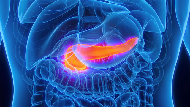
Pancreatic cancer is considered one of the deadliest forms of cancer due to the lack of symptoms during its early stages and the difficulty in detecting tumours before the disease advances.
“The five-year survival rate in the US is about 12 to 13 per cent because of our inability to detect it at a stage where treatment can be most effective,” said Ajit Goenka, radiologist and nuclear medicine specialist at the Mayo Clinic in Rochester, Minnesota.
Researchers noted that while diagnostic methods for many cancers have improved over the years, pancreatic cancer detection has remained a major challenge. By the time tumours become visible through conventional scans and tissue tests, the disease is often already terminal.
The newly developed model, called the Radiomics-based Early Detection Model (REDMOD), uses AI to recognise patterns invisible to the human eye. It converts CT scan images into mathematical representations and analyses the pancreas pixel by pixel after building a three-dimensional image of the organ.
According to Goenka, the system measures subtle differences within the pancreas and compares them with scans of healthy individuals to identify early warning signs.
The research team tested the model on 2,000 previously collected CT scans that were originally taken for unrelated medical conditions. Around one-seventh of those scans belonged to patients who later developed pancreatic cancer.
The AI tool successfully identified 73 per cent of these early-stage cancer cases. On average, the scans analysed by the system had been taken nearly 16 months before the patients received an actual diagnosis.
Researchers said the AI model showed significantly better sensitivity than radiologists in identifying early cancer signs, particularly in scans taken more than two years before diagnosis.
However, experts also noted that the tool still requires refinement. While the AI model identified healthy patients correctly in 81.1 per cent of cases, radiologists achieved an accuracy rate of 92.2 per cent.
Goenka said the technology is intended to support doctors rather than replace them, adding that AI and physician expertise could work together for better outcomes.
The study has received praise from experts in the field. Tatjana Crnogorac-Jurcevic, professor of molecular pathology and biomarkers at Queen Mary University of London, described the findings as “extremely promising”.
She said early detection could transform treatment strategies, especially for high-risk groups such as people with a family history of pancreatic cancer, cancer-related genetic mutations, or new-onset diabetes.
Researchers are currently conducting clinical trials to further validate the model before it can be introduced into routine medical practice. Goenka said the AI system could potentially become part of clinical use within the next five years.
Experts also believe combining AI imaging tools with other diagnostic methods, including urine-based biomarker tests, may further improve the accuracy and sensitivity of early pancreatic cancer detection.