Daijiworld Media Network - Mumbai
Mumbai, Feb 4: A computer-aided detection (CAD) chest X-ray tool has shown diagnostic accuracy comparable to experienced radiologists in identifying pulmonary tuberculosis (TB) in low-resource, high-burden settings, a retrospective pilot study of nearly 500 patients has found.
The study was conducted against the backdrop of limited access to trained radiologists in many TB-endemic regions, where delayed diagnosis can worsen outcomes and increase transmission. Researchers assessed whether CAD-based chest X-ray software could support clinical decision-making in such environments.
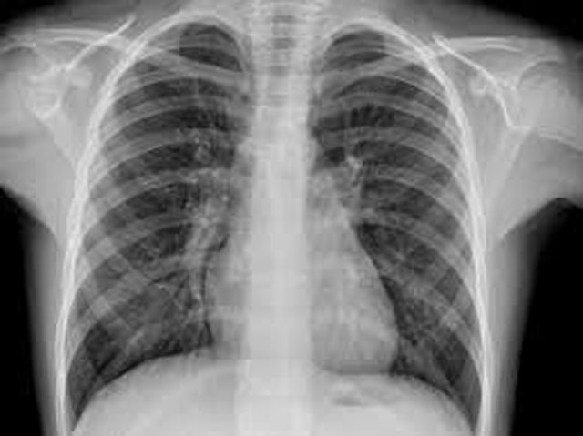
The retrospective analysis included chest X-ray films collected between January 1, 2017 and March 30, 2018 in Guinea-Bissau and Ethiopia. Each image was evaluated by CAD software and independently reviewed by two experienced Ethiopian radiologists. To reflect real-world constraints, the study also analysed images captured by photographing X-ray films using mobile phones and a digital camera.
Final TB diagnosis, based on clinical and laboratory findings including Mycobacterium tuberculosis detection through Xpert MTB/RIF, was used as the reference standard.
A total of 498 chest X-rays from patients with symptoms suggestive of TB were included. Radiologist A flagged 50 images as TB-indicative, radiologist B identified 99, while the CAD software marked 81.
For Xpert-confirmed pulmonary TB, the CAD tool recorded an area under the receiver operating characteristic curve of 0.84. At a cut-off score of 0.5, the software achieved a sensitivity of 76.5% and specificity of 85.9%. Radiologist A showed sensitivity and specificity of 64.7% and 91.9%, while radiologist B recorded 76.5% and 82.3%, respectively.
Agreement on TB-related findings was described as moderate. The combined agreement between the two radiologists stood at κ=0.45, while agreement between each radiologist and the CAD tool was κ=0.56.
Researchers said the findings suggest CAD chest X-ray tools could play a valuable role in supporting TB diagnosis in settings with limited radiology services, even when images are captured using commonly available devices such as smartphones.