Daijiworld Media Network - New York
New York, Nov 22: A combination of computed tomography (CT) and magnetic resonance imaging (MRI) may improve the detection of jawbone invasion in patients with oral squamous cell carcinoma (OSCC), potentially helping doctors plan the extent of surgery, according to a retrospective study.
The study analysed 57 patients with untreated OSCC whose tumours were located close to the mandible. All patients underwent preoperative CT and MRI before surgery between 2016 and 2021. The imaging findings were compared with histopathological examination of the removed jawbone.
Mandibular invasion was confirmed in 25 patients. CT recorded a sensitivity of 88% and specificity of 90.6%, while MRI achieved 92% sensitivity and 87.5% specificity. Both methods had an overall diagnostic accuracy of 89.5%.
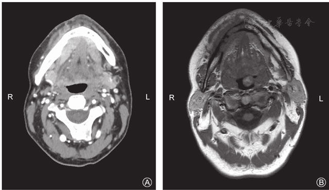
When findings from either scan indicated bone invasion, the combined approach achieved 96% sensitivity and a negative predictive value of 96.4%. However, specificity decreased to 84.4%.
The researchers also examined the detection of cortical and medullary bone involvement. Combined imaging identified cortical invasion with 88% sensitivity and 100% specificity, while sensitivity for medullary invasion was 92%, with specificity of 87.5%.
The higher negative predictive value of combined imaging could help identify patients who may not require extensive jawbone removal, the researchers suggested. However, the study did not establish whether using both scans changed surgical decisions or improved patient outcomes.
The analysis also found differences in five-year overall survival depending on the extent of bone involvement. Survival was 87.4% among patients without mandibular invasion, compared with 55.5% in those with cortical invasion and 37.8% in patients with medullary involvement.
Although the difference was statistically significant, the survival analysis was not adjusted for other factors, including tumour size, lymph node involvement and additional treatment.
The researchers acknowledged limitations, including the retrospective design, small sample size and lack of blinding between radiologists and pathologists.
They concluded that CT and MRI could offer complementary information during preoperative assessment. However, larger studies and prospective research are needed to establish whether combined imaging improves surgical planning and patient outcomes.