Daijiworld Media Network - Turku
Turku, Aug 26: Researchers from the University of Turku in Finland and the Gustave Roussy Institute in France have identified a molecular mechanism that determines whether cells from an aggressive form of colorectal cancer become conventional or inverted as they spread through the body.
The discovery could help identify patients at increased risk of cancer spreading to the abdominal cavity. The study has been published in the journal Nature Communications.
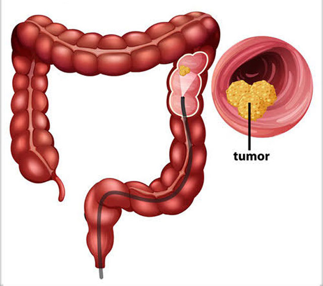
The researchers focused on mucinous colorectal adenocarcinoma, an aggressive form of colorectal cancer that accounts for around 10 to 15 per cent of all colorectal cancers. It is more common among young adults and women.
Unlike most colorectal cancers, mucinous colorectal adenocarcinoma spreads as small, compact clusters of cells known as tumour spheres. These clusters migrate to the abdominal cavity and metastasise on the peritoneum, a pattern associated with poor prognosis and limited treatment options.
Tumour spheres that spread to the peritoneum are typically inverted, meaning they secrete a layer of mucus onto their outer surface. Normally, the mucus is located inside the cell cluster, making it a conventional tumour sphere.
The mucus forms a protective layer around the tumour, reducing the effectiveness of cytostatic drugs while helping cancer cells migrate into the abdominal cavity. The inverted structure also enables tumour cells to invade tissue more effectively and increases their resistance to chemotherapy.
However, when the tumour spheres reach the peritoneum, some revert to their conventional state, with the mucus moving inside the tumour sphere. The cell cluster then attaches firmly to the surrounding tissue, a process believed to promote peritoneal metastasis.
Until now, it had remained unclear what triggers this reversal.
The researchers examined tumour samples from patients and traced the molecular chain reaction that occurs when tumour spheres come into contact with collagen, the structural protein found in the connective tissue surrounding the tumour.
The process results in increased levels of three proteins — SorLA, HER2 and HER3 — in the cancer cells. The activity of these proteins causes the tumour cells to revert to the conventional state and strengthens the attachment of their adhesion receptors, known as integrins, to surrounding tissue.
Analysis of patient samples confirmed the findings observed in laboratory experiments. Levels of SorLA, HER2 and HER3 were highest in tumour spheres in the conventional state, while their levels were lower in inverted tumour spheres.
Since therapeutic antibodies targeting HER2 and HER3 are already available and used to treat other cancers, the researchers investigated whether these drugs could affect tumour-cell inversion and adhesion.
"When tumour spheres grown in the laboratory were treated with antibodies, the cancer cells began to die, were inverted, and their ability to attach to the peritoneum was impaired. The results suggest a potential way of slowing the spread of cancer, but further research and clinical trials are still needed," said Doctoral Researcher Meri Pelkonen from the University of Turku.
Colorectal cancer remains one of the world's leading causes of cancer-related deaths, while its increasing prevalence among young adults has raised growing concern. The reasons behind this trend are not yet fully understood.
Mucinous colorectal adenocarcinoma is more prevalent among younger patients than other forms of colorectal cancer, making a better understanding of its biology particularly important.
According to the researchers, the newly identified mechanism could also explain similar inversion processes observed in other aggressive cancers. It may eventually help identify and potentially treat patients whose tumours are most likely to spread to the abdominal cavity.
The research was funded by the Cancer Foundation Finland, Research Council of Finland, Sigrid Jusélius Foundation and the EU Horizon 2020 programme.