Daijiworld Media Network – Seattle
Seattle, Nov 14: Scientists have created a detailed cellular atlas of the basal ganglia, a key region deep inside the brain that plays an important role in movement, decision-making, emotions and habit formation. The breakthrough could help researchers develop new treatments for several neurological and psychiatric disorders.
The research team mapped different cell types and their gene activity in the basal ganglia using brain tissue from humans, mice, macaque monkeys and marmoset monkeys. Dysfunction in this brain region is associated with conditions including Parkinson's disease, Huntington's disease, schizophrenia, Tourette's syndrome, bipolar disorder, obsessive-compulsive disorder and addiction.
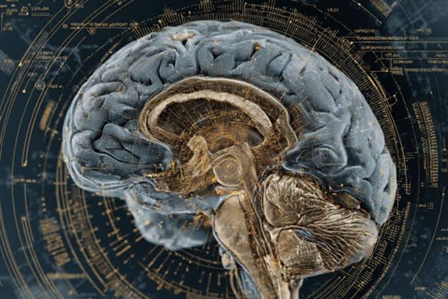
The findings were presented in more than two dozen studies published in the journal Cell and related journals as part of the BRAIN Initiative, launched in 2014 with support from the US National Institutes of Health.
Hongkui Zeng, director of brain science at the Allen Institute in Seattle and one of the research leaders, described the work as an important step towards developing a comprehensive cellular atlas of the entire human brain, targeted for completion around 2028.
Joseph Ecker, a molecular biologist and geneticist at the Salk Institute in California, said there had previously been no comprehensive reference detailing every cell type in the human basal ganglia and how its genes are regulated.
The atlas could initially serve as a valuable reference for researchers studying brain disorders. In the longer term, it may help identify specific targets for gene therapy and gene editing, while allowing scientists to compare diseased brain tissue with healthy tissue. However, treatments based on the findings are likely to take several years to develop.
Among the important findings, researchers identified a previously unknown type of brain cell associated with schizophrenia and bipolar disorder. They also documented genetic changes in specific cell types that could be linked to autism, along with age-related changes in gene activity.
For the study, scientists examined around 21 million cells from human basal ganglia tissue obtained from deceased donors, along with millions of cells from the three animal species.
Margarita Behrens, a neuroscientist and biochemist at the Salk Institute, said comparing the cellular composition across species would help researchers understand brain functions, connectivity patterns and responses to medicines.
The basal ganglia comprise interconnected structures, including the striatum, globus pallidus and substantia nigra. These structures help initiate and regulate movement while also supporting habit formation, reward learning, motivation and emotions.
The atlas combines several layers of information, including genes active in individual cells, chemical and protein modifications that regulate gene activity, and spatial maps showing the location of different cell types within the brain tissue.
Researchers said the information could help identify how genetic variations linked to brain disorders affect specific nerve or support cells. Such understanding could provide an important starting point for identifying disease mechanisms and developing targeted treatments.